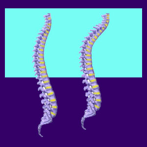
Vertebral wedging is a general term used to describe spinal bones which demonstrate an atypical shape, similar to that of a wedge. This means that instead of being flat with the top and bottom of the vertebral body being basically parallel, the vertebra is instead lower on one side and higher on the other side, forming an asymmetrical trapezium or a triangle shape, instead of a parallel rectangle shape.
Wedging can be easily diagnosed using virtually any form of spinal imaging, including x-ray, CT scan or MRI technologies. The condition can be expressed from incredibly mild to extreme, with a variety of symptoms to be expected ranging from none at all to complete disability.
This essay profiles hemivertebra conditions in the backbone and their potential effects on the form and function of the spine.
Vertebral Wedging Fracture
A vertebral wedge fracture is one possible causes of irregular shaped spinal bones. This type of fracture can occasionally be the result of traumatic injury in any patient, but is far more typical in elderly patients as a compression fracture.
In many cases, wedge fractures in older patients are completely asymptomatic and may go undiagnosed for a long time. In other cases, a wedge fracture can cause bone fragments to enter the central spinal canal, potentially causing spinal stenosis or even spinal cord injury.
In other instances, bone fragments may impinge upon or compress nerve roots in the lateral recess or in the neuroforaminal space, possibly enacting a pinched nerve. Severe wedge fractures have the potential to cause spinal instability.
Wedged Vertebra aka Hemivertebra
Not all wedge shaped vertebrae are due to fracture. Many are congenital or developmental conditions which were apparent from birth or developed as part of the patient’s specific spinal anatomy. This congenital condition is also commonly called a hemivertebra, particularly when the vertebral body is truly underdeveloped and triangular in shape.
Wedged vertebrae are also common in many scoliosis patients and those with advanced forms of abnormal lordosis, kyphosis and are even seen in a few extreme cases of spondylolisthesis.
Wedging can also be misdiagnosed in rare instances when muscular spasms pull vertebral levels very close together. In these instances, preliminary imaging may appear to show a wedged condition, while more advanced imaging will reveal that the actual vertebrae are normal, but the alignment is so out of normal position as to make the spinal bones appear wedged in certain locations.
Vertebral Wedging Facts
I see the notation of wedging listed on many of the MRI reports you send to me for review. In some cases, the findings are part of normal spinal arthritic processes and are minor and inconsequential. In other cases, the findings are certainly cause for concern and should be monitored closely or treated professionally. In a select few cases, the wedging is obviously a big issue and has resulted in an unstable spine which is likely to need drastic surgical intervention to resolve.
As always, I highly recommend speaking to your doctor about any structural finding in your vertebral column and have them explain the results of diagnostic testing in vivid detail. If they can not, will not, or have trouble committing to an opinion, it is time to get a new doctor.


