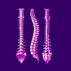
The human spine supports our physical structure and makes possible an incredible and diverse range of motion due to its segmented design. The spine is not only incredibly strong, but it is also flexible, resilient and well engineered for lifelong performance. Each region of the backbone is specifically focused on certain tasks, including support, protection or movement.
The vertebral column is composed of many bones, spinal disc spacers and neurological tissues, but also includes a diversity of soft tissues, such as muscles, ligaments and tendons, holding everything together. Detailed below are all the individual parts of the backbone and information on each component can be found on its dedicated article page.
Vertebral Column Components
The spinal anatomy is made up of 24 individual vertebra, followed by the sacrum and the coccyx. The spinal column may also be called the backbone or the vertebral column. It is important to understand the composition of the spine, especially the lumbar spinal anatomy, since this is the region that seems to trouble most patients chronically.
Vertebrae are the individual bones which make up the spinal column. Each region has its own design, which helps specific vertebral bones to accomplish particular objectives and uses. Vertebrae are thinner and lighter structured at the top of the spine and generally get progressively larger through the end of the lumbar region before tapering back off at the tail end of the spinal column.
Intervertebral discs are natural shock absorbers that cushion the space in between individual vertebral bones. Discs are made of water, collagen and proteoglycans. Discs facilitate individual vertebral level movement and provide for an overall flexible spine.
Annulus fibrosus is the woven layered outer wall of the spinal disc. This outer disc wall is very tough, but will almost inevitably suffer degeneration with age, especially in particular locations which endure the most movement. Annular tears can allow the nucleus of the intervertebral disc to extrude into the greater spinal anatomy.
Nucleus pulposus is the soft inner core of the spinal disc structure. This moisture-rich gelatinous nucleus is what gives the disc its shock absorbing qualities.
Thecal sac is the protective membrane surrounding the spinal neurological tissues. Learn how thecal sac impingement might cause back or neck symptoms, but can also be an incidental diagnostic finding.
Foramen, also called neuroforamen, are the openings in between vertebral bones through which the spinal nerves exit at each level of the backbone. When these suffer stenosis, the blockages are often implicated in causing pinched nerves.
Spinal cord is the central nerve structure which serves as a pathway for messages between the brain and the body. The spinal cord also is responsible for carrying out many of the body’s reflex actions, completely independent of the brain.
Cauda equina is the bundle of spinal nerves which continues through the lumbar, sacral and coccygeal vertebrae at the end of the spinal cord.
Facet joints exist on the posterior of the vertebrae and help provide intervertebral links. These synovial joints join the individual spinal bones to the vertebrae above and below.
Nerve roots are the neurological tissues which branch off the spinal cord, forming the network of nerves which run throughout the body. Nerve roots exit at each spinal level bilaterally.
Spinal nerves serve the neurological requirements of the entire anatomy.
Sciatic nerve, also called the ischiatic nerve, is the largest and one of the most important nerves in the body.
Brachial plexus is a concentrated grouping of neurological and vascular tissue in the upper back and neck.
Back muscles help to support and stabilize the spinal structures. The muscles of the back are some of the strongest in the entire body and are well designed to provide us with the abilities to do all the physical tasks we require during our lifetimes.
Cervical spinal region is the first 7 vertebrae in the spinal column, also called the neck. Read all about the complex cervical spinal anatomy.
Thoracic spinal region is the 12 vertebrae between the cervical and lumbar areas, often called the upper back or middle back.
Thoracolumbar region is the frontier between the middle back and lower back.
Lumbar spinal region is the final 5 vertebrae in between the thoracic area and the sacrum, commonly called the lower back.
Lumbosacral region is the juncture between the lower back and the sacrum.
Sacrum is 5 fused vertebrae in between the lumbar area and the coccyx.
Coccyx is 4 fused vertebrae in the lowest area of the spinal column, also known as the tailbone.
The epidural space houses many of the vital spinal structures, including components of the central nervous system.
Schmorl’s nodes are a common vertebral/intervertebral abnormality.
Chiari malformation is a potentially catastrophic abnormality of the foramen magnum known to contribute to spinal syrinx formation.
Transitional vertebra usually occur at the lumbosacral juncture, but can also exist at the cervicothoracic junction, as well.
Hemivertebra can exist anywhere in the spine and might have significant functional and structural consequences.
Sacralized vertebra are naturally fused lumbar bones sometimes blamed for enacting pain.
Spinal cysts can form on or near the backbone or spinal cord.
Ligamentum flavum is one of the crucial spinal ligaments often implicated in causing pain syndromes which are typically diagnosed as ligamentum flavum thickening or hypertrophy.
Longitudinal ligaments help to regulate movement and stabilize structure of the vertebral column. Meanwhile, ossification of the posterior longitudinal ligament is one of the more problematic of the age-related degenerative changes to the backbone.
Naturally fused vertebrae can form almost anywhere in the backbone, due to injury, degeneration or congenital issues.
Learn more about normal and abnormal vertebral column anatomies in our article detailing spinal curvature.
The Wonderful Spine
The human spinal column is a true miracle of design and functionality. Although implicated in a wide range of back pain conditions, the backbone is only sometimes the actual source of ongoing severe symptoms. It is crucial to remember that age will create degenerative processes in the spinal column. However, these processes are normal and not usually the source of significant pain or any major health concerns in their normal mild to moderate expressions.
Learn all about spinal degeneration and how it is blamed for causing back, neck and sciatica pain.
Read more about how to best utilize spinal diagrams when researching your diagnosis.
Have you been diagnosed with a spinal deformity? If so, take the time to learn more about your specific abnormal structural or functional condition.
Learn how to maintain a healthy vertebral column for life with some simple guidance from our editorial team.


