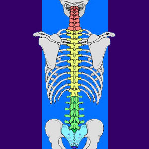
Studying a spine diagram is one way to better understand many of the individual components of the back bone and how they might relate to a symptomatic back, neck or sciatica pain condition. So many patients receive diagnostic imaging reports full of terms and anatomical locations which are unknown and mysterious to them. Many of these patients take to the internet in order to find out additional information, since it is rare that a physician or chiropractor will actually spend adequate time explaining the structural findings in the detail they deserve.
This article provides some guidance for patients who are looking to clarify their back pain diagnosis using spinal pictures, photos and diagrams.
Spine Diagram Components
When looking at the human spine, it is crucial to understand that there are many different types of tissues which make up the structure:
Spinal bones, called vertebrae, are often location-specific in shape and function. No 2 vertebrae are exactly alike in size, shape or functionality,
Intervertebral discs are the soft cushiony spacers in between most vertebrae. There are spinal discs in between all typical vertebral bones, with the exception of the first two, called the atlas and the axis. Disc spacers are also absent in between the naturally fused bones of the sacrum.
The spinal cord in the central conduit of life energy, linking the brain and the body. This cord follows the natural contours of the vertebral column and eventually separates in the upper lumbar region to form the cauda equina.
Spinal nerve roots branch off from the spinal cord and exit the central canal at every vertebral level in order to create the network of nerves which serve the needs of the entire body.
There are also less commonly known parts of the spine, including major and minor blood vessels, ligaments, muscles and a variety of protective and encapsulating membranes.
Spine Diagram Relevance
I find the following to be relevant points when discussing diagrams of the spine, as they are used by most back and neck pain sufferers:
People often have a difficult time placing the location shown in a picture of the spine into their actual body. Where exactly are these structures located? It may be impossible to tell without the skin or other anatomical markings superimposed on the diagram.
Are the diagnosed structural issues causative or contributory? Spinal abnormalities are commonplace and often quite innocent, particularly when there is unresponsive chronic pain or a set of symptoms which does not correlate to the anatomical findings. Are spinal abnormalities located during diagnosis really problematic or just anatomical differences? Most atypical spinal anatomies are not concerns, although some may be symptomatic in extreme cases.
Is spinal degeneration a real threat to health or is it just part of the normal and expected aging process? In most cases, normal degeneration is nothing to fear.
Spinal Conclusions
Go ahead, study the spine. I do. It will not hurt and it just might help you to better understand any diagnostic verdict which you may have received. Knowledge is always a good thing.
Just be sure to understand that just because your spine does not exactly mirror a perfect specimen, this does not necessarily explain any back, neck or sciatica symptoms. In fact, just try and find an actual example of that perfect spine in a living human. You will be hard pressed. Degeneration and structural irregularities are the rule, not the exception.
You will likely have these atypical conditions and so does your doctor, as well as virtually everyone else on the planet. Learn the facts before you blame normal structural deterioration for pain, particularly when the symptoms may not support the diagnostic theory.


