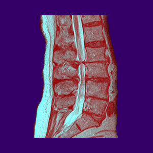
A narrowed spinal canal, also called spinal stenosis, is a commonly reported result on many MRI reports and can be a confusing and worrisome finding for any affected patient. The spinal canal is the space inside the vertebrae containing the spinal cord, the spinal nerve roots, some blood vessels and a variety of membranes which protect these structures.
There are many reasons why the spinal canal, also called the central canal, may be narrowed and some of these conditions may create the ideal circumstances for back pain and related neurological concerns. This treatise provides details of these origins of stenosis and how the contributors often act together to slowly reduce the overall patency of the central canal.
What is a Narrowed Spinal Canal?
The spinal canal is technically the space inside the vertebrae (also called the vertebral foramen) containing the neurological tissue. However, most doctors and patients alike use the term to describe the actual soft tissues inside the canal itself. These tissues include the various levels of meninges (dura mater, arachnoid mater, pia mater), the spinal cord and the spinal nerve roots. The thecal sac is the name for the membrane which protects the nerve tissues and is filled with cerebral spinal fluid.
This central canal has extra space to ensure that the spinal cord and nerve roots have plenty of room to facilitate proper neurological function. Therefore, a normal spinal canal is not compressed or impinged upon in any way. In a narrowed central canal, the diameter of the space is reduced, typically in a specific area of the structure, possibly causing some neurological concerns in extreme cases.
Causes of Spinal Canal Narrowing
There are many possible reasons for narrowing of the central canal. It is completely normal and expected that people will endure narrowing as they age, since most of the typical sources of canal narrowing are universal parts of getting older:
Congenitally narrowed canals are less patent than typical from birth. This may be demonstrated in specific areas or within the entire canal space in rare cases.
Prolapsed discs can impinge upon the central canal, often noted as thecal sac impingement or compression on MRI reports. This is very, very common and almost always a non-issue. If the disc compresses the spinal cord or cauda equina, the patient has a much greater chance of suffering related symptoms.
Ligamentum flavum hypertrophy might worsen the effects of a central herniated disc or might work to create a stenotic effect on the posterior of the canal space in combination with any other anterior narrowing issue.
Arthritis in the spine is normal and will cause narrowing of the central canal due to the build up of arthritic debris, osteophytes and osteochondral bars.
Spondylolisthesis and other vertebral misalignment conditions can narrow the central canal or cause it to not line up correctly, possibly compressing the spinal cord or nerves.
Scoliosis and other abnormal spinal curvatures, such as lordosis or kyphosis, can narrow the central spinal canal and trap nerves or impinge on the spinal cord.
Fractured vertebrae can narrow the spinal canal due to acute trauma or compression fracture events.
Narrowed Spinal Canal Consequences
A narrowed central canal is a usual finding in patients over the age of 50, although it can occur far earlier. It most often occurs in the middle to lower cervical spine and in the lower lumbar levels and lumbosacral juncture. The condition may be caused by unspecified arthritic change, including bone spurs combined with disc desiccation. The condition may be worsened or sourced by a specific pathology, such as a herniated disc or spinal alignment condition. Regardless, the finding of a narrowed canal is rarely a reason for worry, unless there is such an extreme narrowing that the spinal nerve structures do not have room to operate correctly or there is no circulation of cerebral spinal fluid. These events are rare, but they do occur.
Notation of mass effect, impingement, displacement or effacement of the spinal cord or nerve roots does not guarantee symptoms. Notation of compression of the spinal cord or nerve roots increases the chances for symptoms drastically.
It is vital to understand the basic spinal anatomy in order to better your chances of preventing misdiagnosis and therefore, unsuccessful treatment. The spine can be complex, but with a bit of knowledge and a terrific tool like spinal MRI, patients can truly understand why they may have pain, as well as the comprehend structural conditions which may exist, but are not in any way symptomatic.


